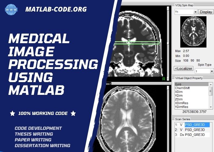
This article is about the fundamental information of medical image processing using Matlab tool with its applications and techniques. Medical imaging means acquiring or creating medical images of human body parts through intelligent techniques for medical commitments. The main intention of this technique is to help the physician detect and treat the clinical disease of the patients. For once-a-week, infinite numbers of imaging processes are globally performed.
The increasing growth of image processing eventually increases medical image processing advancement through their incredibly accurate image processing techniques such as image segmentation, detection, analysis, classification, enhancement, and visualization.
The degree of tissues detection is gradually growing due to the advanced image processing approaches. This process comprises radiological imaging, which utilizes electromagnetic energies (gamma and X-rays), organic, magnetic, sonography, isotope, and thermal imaging.

Medical Image Processing Using Matlab
Here, we have given other major medical image processing and analysis functions for your reference.
- Edge Enhancement
- Local Spectral Analysis
- Median Filtering (3D)
- Image Arithmetic
- Contrast Enhancement
- Convolution
- Morphological Transforms
- Hilbert Transform (2D)
- Fast Fourier Transform (FFT)
- Geometric Transformations
- Finite Impulse Response (FIR) Filters
- Histogram Equalization (Processing)
- Color Transformation (8 / 24 bits)
- Wavelet Transforms (Continuous and Discrete)
- High Pass Filtering and also Low Pass Filtering
- Region-of-Interest (RoI) Extraction and Selection
Next, have a look at common medical image types that are commonly used in Medical Image Processing using Matlab. These images are acquired or created differently from different medical equipment sources by using various imaging techniques. Each image represents any of the organs, tissue, and muscle of the human body.
Medical Image Modalities
- Ultra-Sound (US)
- Cranial, Abdominal, Breast, Spleen, Doppker, Gallbladder, and others
- CT
- Brain, Chest, Cervix, Breast, Kidney, Pancreas, Lungs, Bladder, Chest, Esophagus
- And more
- MRI
- Gastrointestinal Tract (GT), Cardiovascular, Oncology and Liver Images
- Hybrid Modality
- US with CT, PET with MRI, US with MRI, MRI with CT, SPECT with CT, PET with CT, MRI with SPECT, etc.
- X-Ray
- Discography, Oncology, Upper GI, Neuro Imaging, Cardiology Images, Arthography, Pharmacokinetics, Dexa Scan, Fluroscopy, Infected Tissues, Mammography Images, Contrast Radiography , Positron Emission Tomography (PET) Scan and also many more.
Next, some of the important real-world applications of medical image processing are given for your information. All these applications use the image dataset as input and implement appropriate image processing based on application requirements.
List of Applications
- Real-Time Cancer Prediction and Detection
- Enhanced Image Filtering in Pattern Recognition
- Fast Medical Image Retrieval in Healthcare Applications
- Medical Image Fusion in Remote Sensing Applications
- Breast Cancer Detection and Screening using Mammograms
- Efficient Image Denoising in Real-Time Document Processing
- Improved Medical Image Compression in Telemedicine Applications
As a matter of fact, Matlab is the best tool for medical image processing, where you can find the sophisticated infrastructure for implementing all medical image processing techniques. Through this platform, you can easily access, process, analyze and view medical data (signals/images). Also, it enables the developers to build, test and deploy new algorithms and mechanisms.
In the coming section, you will see the importance of the need of the MATLAB and Toolboxes with their bundled software and functions. Overall, it is an efficient mathematical tool to get the desired results from MRI, PET, Fluorescein Angiogram, and CT images. In specific, we have given the reason behind the use of Matlab in medical image processing.
Why to use Matlab for Medical Image Processing?
- Easy to automatically create the label the image data in the folder tags
- Scalable to collect the large data from multiple sources and store in memory
- Easy to load the image datasets for rapid processing in real-time and non-real-time applications of computer vision and machine learning
Further, we have given the fundamental operations in processing medical images. Each process has a certain task and goal to achieve. All these processes are collectively combined together in the project to create medical applications for diagnosis purposes.
Operations in Medical Image Processing
- Preprocessing (Denoising and Filtering)
- Segmentation
- Extraction (Features)
- Recognition
- Enhancement
- Reconstruction
- Morphological Operations
- Classification
As mentioned earlier, medical image analysis deals with a large volume of data.
For instance: measuring a patient’s health through wearable devices. Although the data is large, it accurately gets the results by applying advanced methods and algorithms.
Next, we have selected “segmentation” as the sampling process for demonstration. The major process in medical image processing is segmentation. It precisely and automatically delimitates the objects in the time of recognition in spite of data size. In dealing with the subject of the medical process, it segments the brain tumors, blood vessels, liver, left ventricle, etc., from medical images. Further, we have also listed the few methods specifically designed for segmentation purposes.
List of Current Medical Segmentation Methods
- Bayesian Filtering
- HeadLocNet
- Neuro Fuzzy
- Deep Atlas Network
- K-means
- Multi-Thresholding
- Fuzzy
- Active Contour Snake Model
- Diffusion Weighted Model
- Adaptive Sparsity based Algorithm
- Dense Inceptioned U-Net
- Augmented Stain Deep Convolutional Neural Network (D-CNN)
What are the feature extraction methods?
Similar to segmentation, feature extraction is also an important task to be performed in image processing. It helps to extract the essential features from the whole dataset by eliminating redundant data, unwanted data, corrupted data, and irrelevant data.
If you give the whole dataset as input before the feature extraction process, then the processing time will increase on applying the Algorithm on redundant data and others. The data is reduced into a set of features based on the project needs to be called feature extraction to avoid this issue. To improve the accuracy, it is again reduced by selecting the optimal features called feature selection. Now, the processing time will decrease and increase the result precision. Below, we have given the list of algorithms that are used for feature extraction and selection purposes.
- Isomap
- Latent Semantic Analysis (LSA)
- Partial Least Squares (PLS)
- Non-linear Dimensionality Reduction
- Principal component analysis (PCA)
- Kernel PCA
- Independent Component Analysis (ICA)
- Multifactor Dimensionality Reduction (MDR)
Next, we can see the hand-crafted feature concept. It means extracting the properties using different mechanisms in the own image data. Ultimately, it is used to represent the image in the new dimension of perception. Below, we listed some techniques used for Hand Crafted Feature Representation.
Hand Crafted Feature Representation
- Block based Feature Solution
- Intensity based
- Moment based
- Frequency based
- Dimensionality based
- Important Feature Points based Solution
- Speeded Up Robust Features (SURF)
- Harris Corner
- Scale-Invariant Feature Transform (SIFT)
At the present time, deep learning models are widely used in the majority of Medical Image Processing using Matlab. Since deep learning algorithms have several in-built beneficial capabilities to perform any complex image analysis problem in a simplified way, they also work fast and efficiently to yield accurate learning results. So, it attains an inseparable place in the field of digital pathology. For instance: we have given the deep learning-based different image analysis processes involved in the medical application.
- Mitosis and Lymphocytes Detection
- Nuclei and Glands Instance Segmentation
- Healthy and Cancer Tissue Classification
In the current scenario of the COVID-19 pandemic, radiologists suggest MRI as an add-on modality to view and study the long-term COVID-19 characteristics. For this kind of situation, deep neural networks (DNN) play a significant role in analyzing the underlying features of COVID-19 disease.
In general, DNN is the part of the Artificial Neural Network (ANN) which there is an increased number of deep layers, units/layers, and training metrics for processing. Though it has a complex structure, it works effectively than others in terms of accuracy.
Matlab has a special separate toolbox for deep learning models called Deep Learning Toolbox. It offers a well-refined environment to design and implement DNN algorithms, apps/software, and pretrained models. For instance, below, we have given the algorithms used for data classification and regression.
- Input: Text data / Time-Series data / Image
- Aim: Regression and Classification
- Approaches
- Long Short-Term Memory (LSTM)
- Convolutional Neural Networks (CNNs)
Now, we can see the implementation process of the deep learning networks using the deep learning toolbox supported by the Matlab tool. Here, we classify the process into three following modules.

How does Deep Learning Toolbox Works?
- Study of Deep Learning Networks
- Prior to training, explore the network structural design completely for detecting layer compatibility problems and bug fixing.
- Next, view the network structure for finding the information about the learnable activation and parameters
- Design of Deep Learning Networks
- Utilize the Deep Network Designer app for developing deep network from scratch and train them
- For that, first import already trained model and thoroughly view the network architecture. After that, tune the parameters and modify the layers for training
- Management of Deep Learning Trials
- Utilize Experiment Manager app for controlling different deep learning experiments
- For that, it monitors the various trials’ results, code and training parameters
- Further, employ the visualization tools for exploring confusion matrix, classifying plots, assessing trained models and filtering trial outcomes
In addition, we have given how the data is trained in the deep neural network system. And, it specifically explains the deep transfer learning algorithm and deep network designing through training data, training time, computation, and model accuracy.
How the Deep Neural Network Model is trained?
- Training Data
- In performing deep transfer learning based on pre-trained models, it uses labeled images in range of 100-1000
- In creating a new deep network, it uses labeled images in range of 100 – 1,000,000
- Training Time
- In performing deep transfer learning based on pre-trained models, it takes seconds to minutes time for execution
- In creating a new deep network, it takes days to weeks for execution
- Computation
- In performing deep transfer learning based on pre-trained models, it is moderately intensive (GPU optional)
- In creating a new deep network, it is computationally intensive (GPU require)
- Model Accuracy
- In performing deep transfer learning based on pre-trained models, it has decent accuracy which based on pre-trained model
- In creating a new deep network, it has greater accuracy, but it is over fitting to the small data sets
Our research team has given you a sequence of newly born deep learning algorithms that are highly used in medical image processing using matlab for your benefit. Currently, we are working on the below-specified approaches for advanced developments.
Latest Deep Learning Algorithms for Medical Image Processing
- Dynamical Language Model using Meta-Learning Approach
- Few-shot Learning based on Optimization Model
- Siamese Neural Networks for Disorder Severity Detection
- Nonparametric Hierarchical Bayesian Model based on One-shot Learning
- Large-scale Diffusion Simulation using Low-shot Learning Model
- Efficient Deep Regularization Learning and K-Shot Classification
- Semi-supervised Few-shot Learning Classification using Meta Learning
- Few-Shot and Meta Learning Methods for Fast Learning
- GAN based Residual Factor Analysis in Pairwise Networks for One-shot Learning
- Hallucinating and Shrinking Features based Low-shot Visual Object Recognition Prototype Model
Similarly, a medical image processing toolbox is used in MATLAB.
Matlab has various toolboxes which have a colossal collection of packages to support all sorts of medical image processing techniques. Further, this package encloses a set of predefined functions and classes which to make the medical image operations at ease. It offers evidently proved in-built methods for coping with image orientation, rotation, origin, spacing, and many more. Also, this package contains primary image processing processes, input/output functions with different image and mesh formats. Further, we have listed some key functions that are most important for implementing Medical Image Processing projects using Matlab.
Medical Image Processing Toolbox Functions
- MedicalImageProcessingToolbox/processing/Misc
- cropImage
- sphereRandomPoint
- angleInRange
- MedicalImageProcessingToolbox/processing/Sources
- sphereMesh
- boxMesh
- sphereSquarePatch
- coneImage
- prismMesh
- cylinderMesh
- meshPatch
- planeMesh
- circle3DMesh
- ellipsoidMesh
- prismImage
- cylinderImage
- planeImage
- MedicalImageProcessingToolbox/class_mesh
- MeshType
- QuadTetMeshType
- AttributeType
- QuadMeshType
- MedicalImageProcessingToolbox/processing/IO
- write_vtkMesh
- read_roi
- read_FacetTessFile
- writeDOFToFile
- read_parrec2DFlow
- read_offMesh
- write_rec
- readpar
- read_vtkMesh
- readrec
- read_meshMesh
- read_FacetMesh
- write_gipl
- read_gipl
- read_stlMesh
- write_ITKMatrix
- read_gipl2
- read_ITKMatrix
- write_gridAsVTKMesh
- read_CHeartMesh
- read_vtkSP
- read_mhd
- write_CubitMesh
- read_nifty
- write_stlMesh
- read_CubitMesh
- read_parrec
- write_mhd
- read_MITKPoints
- MedicalImageProcessingToolbox/class_image
- VectorImageType
- ImageType
- VectorPatchType
- PatchType
- MedicalImageProcessingToolbox/processing/Geometry
- quadraticX
- intersectionCircleCircle
- dQuadratic
- intersectionCircleFrustum
- barycentricCoordinates
- intersectionPlanePlane
- intersectionLineLine
- PointInFrustum
- intersectionLineSphere
- PointInTriangle
- vtkMathPerpendiculars
- PointInTriangle3D
- intersectionCircleTriangle
- quadratic2D
- intersectionTriangleTriangle
- quadraticValue
- intersect_frustumSphere
- quatToRMat
- intersectionSphereSphere
- quatToRMat_igtl
- intersect_meshSphere
- MedicalImageProcessingToolbox/processing/Visu
- plotAxis
- viewMesh
- plotpoints
- plotboundingbox
- plotpoints2
- MedicalImageProcessingToolbox/processing/Basic
- resliceImage
- gaussND
- gradientImage
- resampleImage
- setOrientation
- bilateralFilterImage
- localMinimaImage
- transformImage
- matrixToRGB
- gaussianBlurImage
- gradientBinaryImage
- differenceOfGaussiansGradientImage
- MedicalImageProcessingToolbox/processing/Meshes
- csgMeshes
- closeMeshWithPlanes
- meshClip
- meshConditionalDilate
- angleSort3d
- imageToMesh
- AABB_node
- meshConditionalErode
- combineMeshes
- meshBinarize
- csgSpheres
- distanceMeshToMesh
- meshFillHoles
- AABB_tree
- intersectEdgePlane
- meshTransform
- meshDilate
- surfaceFlattening
- meshErode
- transformPointsBetweenMeshes
- meshReconstruct
- resample_lines_mesh
- surfaceFlatteningConstrained
- meshToImage
- intersectionSpheremeshPlane
- meshIntersectImage
Finally, yet importantly, our research team likes to let you know the latest research topics on medical image processing. Beyond this, we have numerous research areas and ideas for active research scholars.
Research Ideas in Medical Image Processing
- Advance Techniques for MRI Data Volume Rendering and Visualization
- Efficient Automatic Blood Vessel Tortuosity Measurement
- CNN-based Video Fluorescein Angiogram Analysis and Classification
- 3D Volume Creation from 2D Image Visualization
- Huge-Scale Multi-Resolution Processing on High-Resolution Images
Further, if you want more information about implementing medical image processing using matlab, just make a bond with us. Our experts will fulfil your research requirements in a timely manner through our best research and development guidance.


